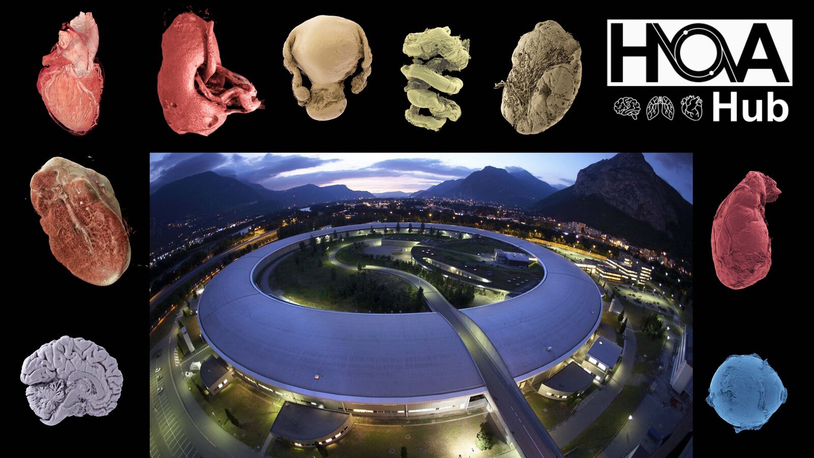


An international team of scientists has announced in the journal Science Advances the launch of a new 3D portal that allows users to explore intact human organs in unprecedented detail – from the entire organ down to individual cells. The Human Organ Atlas (HOA), developed with the participation of the Institute of Pathology at Uniklinik RWTH Aachen, is freely accessible and offers a new way of understanding human anatomy and diseases.
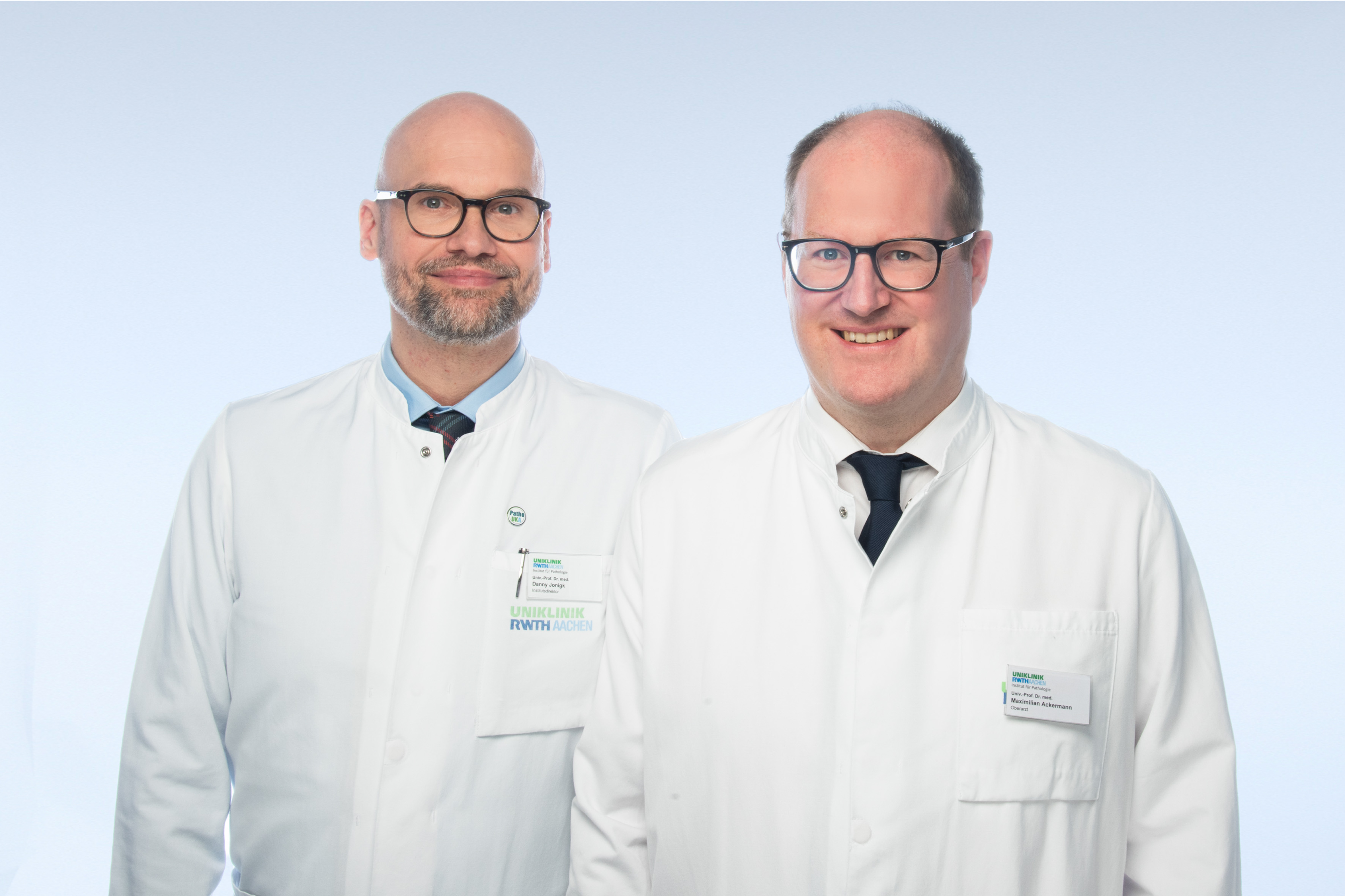
Building on an initial version, the Human Organ Atlas (HOA) is now available in a greatly expanded form and can be accessed directly via a standard web browser without the need for special software. The atlas is based on an advanced imaging method called Hierarchical Phase-Contrast Tomography (HiP-CT), which was developed at the European Synchrotron Radiation Facility (ESRF) in Grenoble, France, by an international team led by University College London (UCL). Prof. Danny Jonigk, Head of the Institute of Pathology at Uniklinik RWTH Aachen, and his colleague Prof. Maximilian Ackermann were also involved in its development.
HiP-CT uses the Extremely Brilliant Source (EBS), a synchrotron source that is up to 100 billion times brighter than conventional CT scanners in hospitals. This allows researchers to scan entire intact human organs non-destructively and then zoom in to near-cellular resolution. ‘To our knowledge, the HOA is currently the highest-resolution open 3D dataset of intact human organs and thus represents a significant advance in biomedical imaging,’ says Prof. Jonigk.
A unique tool for AI, medicine and education
Beyond promoting anatomical and biomedical research, the atlas is set to become an important resource for artificial intelligence. Large, high-quality 3D datasets are rare, which limits the development of advanced medical AI systems. The Human Organ Atlas offers a curated, hierarchical dataset that is ideal for training machine learning models for segmentation, disease detection and super-resolution analysis.
Students can also use the platform as an immersive alternative to traditional anatomy diagrams to build a clearer spatial understanding of complex structures. The research team plans to expand the collection in the coming years and add more organs, samples and new tools. The long-term goal is for the atlas to provide new insights into the internal architecture of the human body, thereby promoting research, education, AI development, medical understanding and public interest in science.
‘This data could fundamentally change the way anatomy is studied and understood,’ emphasizes Prof. Ackermann. ‘I look forward to seeing what else we can achieve in the coming years.’
You can access the open 3D portal here: https://human-organ-atlas.esrf.eu