
Open positions at MOCA
Im Institut für Molekulare und Zelluläre Anatomie der Uniklinik RWTH Aachen sind zum frühestmöglichen Zeitpunkt folgende Stellen zu besetzen:
The Institute for Molecular and Cellular Anatomy (MOCA) is seeking a
Master student (f/m/d) or Medical student (f/m/d) for the project
“Role of intestinal intermediate filaments in the microbial stress response”
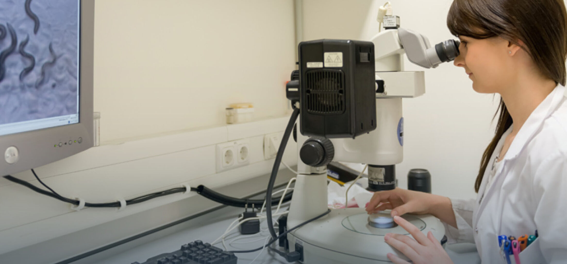
The project investigates the role of the intestinal intermediate filament cytoskeleton in microbiome-host interactions. It exploits the easy handling of the commonly used model organism Caenorhabditis elegans to explore underlying genetic and molecular mechanisms. A panel of well-characterized C. elegans mutants is available to study the responses to defined microbes.
Project aims:
To elucidate the role of the intestinal intermediate filaments in the microbial response we examine
- how intermediate filament dysfunction affects microbial colonization, microbe-induced epithelial reactions and animal health
- how microbiota affect intermediate filament expression
- which pathways are implicated
Your tasks:
You will
- perform microbiome confrontation experiments
- quantify intestinal colonization
- evaluate epithelial physiology
- investigate the innate immune response
- analyze intermediate filament expression
Your profile:
You
- are highly motivated to perform wet lab work
- have a keen interest in genetics, molecular biology and microscopy
- are an above average student in biology, biotechnology or a related discipline with a bachelor degree or an advanced medical student
We offer:
- A research topic with high medical relevance
- Dedicated and tight supervision
- Financial support
If you are interested, send a short letter of motivation, your CV and transcripts to fgeislerukaachende .
Jun.-Prof. Dr. Florian Geisler
Institute for Molecular and Cellular Anatomy
Wendlingweg 2
RWTH Aachen University
D-52057Aachen
fgeislerukaachende
https://www.moca.rwth-aachen.de/c_elegans.html
We are currently seeking a highly motivated
life science student (f/m/d) for a master thesis on
“Mechanical homeostasis of retinal pigment epithelium in ageing”
Age-related macular degeneration is the most impactful blinding disease of the elderly population. The most vulnerable outer retinal layer involved in AMD pathogenesis is the retinal pigment epithelium (RPE). RPE cells experience severe remodelling during the disease - alterations of their extracellular matrix, cellular hypertrophy, increased size heterogeneity and severe cytoskeletal remodelling. These alterations strongly suggest changes in epithelial mechanics. With age being the major risk factor for AMD, it is crucial to understand how the healthy RPE ages in terms of mechanics to identify when a phenotypic switch may lead to AMD.
The RPE is a post-mitotic epithelium, meaning cells cannot proliferate in response to apoptosis. Cellular hypertrophy and reconfiguration compensate for the linear reduction of RPE cell numbers with age. The induction of apoptosis in vitro can mimic RPE ageing and provide the opportunity to ‘mechanically age’ the epithelial monolayer similarly to material ageing. It is unknown if this age-related density reduction affects monolayer mechanics of the RPE and what implications the effects have on RPE function.
Project aim:
The project aims to characterise epithelial mechanics of an ageing post-mitotic epithelium. Ageing of hiPSCs-derived RPE cells will be mimicked by inducing large-scale density reduction. At the same time, mechanics will be analysed in terms of traction forces, stresses within the monolayer, cell stiffness and monolayer arrangement.
If you are interested, please send a short motivational letter, CV and transcripts at jdirussoukaachende
We offer:
- Interdisciplinary and active research environment
- Close practical and theoretical supervision
- Possibility for ending up in publication
Your tasks:
- Culture of hiPSCs-derived RPE on polyacrylamide hydrogels and ageing-mimicking stimulation
- Traction force and monolayer stress microscopy of ‘aged’ vs control cells
- Segmentation and image analysis
- Optional: Nanoindentation, immunofluorescent staining
Your profile:
- Master student in Biology, Biotechnology, Biomedical engineering, or a related discipline
- Motivated, focused and team-oriented attitude
- Experience with cell culture, microscopy, AFM, Fiji or Matlab are a plus
If you are interested, please send a short motivational letter, CV and transcripts at jdirussoukaachende.
We are currently seeking a highly motivated
Master student (f/m/d)
for a project on “ Regulation of cardiogenesis in desmosome deficient mouse embryos”.
Desmosomes are important structural junctions between cardiomyocytes that maintain cellular structure and confer tissue integrity. In the “Heart research group” of MOCA we aim to study the impact of mutations in desmosomal proteins during early heart morphogenesis. To this end, transgenic animal models will be utilized to investigate in vivo cardiogenesis under specific genetic conditions. In the first phase of the project, desmosome formation will be compared in different transgenic animals to wild type controls. In the second phase of the project cardiomyocytes shape, cytoarchitechture and their extracellular matrix composition will be studied.
Your Tasks
- Histological analysis of embryonic heart, including tissue fixation, sectioning by microtome and staining of the tissue slides
- Detection of protein expression and localization using immunohistochemical techniques
- Visualizing protein expression and localization by fluorescence microscopy and analyzing data via ImageJ software.
- Analysis of RNA expression and localization via in situ hybridization techniques
Your profile
- B.Sc. in biology, biomedical or related studies
- Interested in developmental aspects of heart
- Previous lab experiences is a plus but not required
- Detail oriented, a good observer, and well organized
Please send your application including your CV, cover letter and transcripts to Mrs. Dr. Hoda Moazzen, hmoazzenukaachende.
Institute of Molecular and Cellular Anatomy
RWTH Aachen University
Wendlingweg 2
D-52057 Aachen
Phone: +49 (0) 241 80 85298
Email: hmoazzenukaachende
Web: https://www.moca.rwth-aachen.de/heart_disease.html
We are currently seeking a highly motivated
medical student (f/m/d)
for a project on “Mechanobiology of genetically modified retinal epithelium”.
The integrity and homeostasis of the retinal pigment epithelium (RPE) are critical to sustain the healthy function of the retina. RPE cells tightly interact with each other, forming a monolayer of cuboidal and polarized cells located between the choriocapillaris and the photoreceptor outer segments. It fulfils multiple tasks, including the maintenance of the blood-retina barrier to protect the retina, the transport of nutrients, the removal of metabolic products and the secretion of vital factors and molecules. Degeneration of the RPE interferes with the normal retinal metabolism, breaks the blood-retina barrier and causes vision loss. The RPE undergoes chronic mechanical stress, which generally plays a critical role in the (patho-)physiology of living cells. Dysfunctions contribute to the pathogenesis of many retinal degenerative diseases, such as proliferative diabetic retinopathy (PDR), proliferative vitreoretinopathy (PVR), RPE tears and high myopia. These diseases have global prevalences of up to 3% and are characterized by harmful stretching of the RPE, resulting in the distortion of the retinal architecture up to retinal detachment and leading to the disruption of the essential interactions between RPE and outer retina and, finally, to blindness.
This project aims to characterize the mechanobiology of genetically modified retinal pigment epithelium. The genes for pigment epithelium-derived factor (PEDF) and brain-derived neurotrophic factor (BDNF) are delivered via electroporation using the Sleeping Beauty transposon system. The functionality of the monolayers from non-transfected and transfected cells will be performed by electrical impedance spectroscopy to monitor RPE barrier properties and morphological changes, as well as traction force microscopy and monolayer stress microscopy to evaluate RPE mechanobiological properties.
The resulting characterization of the genetically modified monolayers will set a step forward for the use of the transfection strategy in a novel therapeutic strategies.
Your Tasks
• Optimization of traction force microscopy and electrical impedance spectroscopy of RPE.
• Live imaging of RPE monolayer’s actin cytoskeleton with confocal microscopy
• Computational data analyses using MATLAB and Fiji software
Your profile
• You are studying medicine
• You are interested in the field of mechanobiology
• You are interested in mammalian cell culture
• You are a reliable and careful worker with the ability of integrating well in a team
The student will have the possibility to apply for a scholarship to the German Society of Ophthalmology and will be working in a team of engineers, physicists, biologists and medical doctors from the groups of Dr. Di Russo (UKA, DWI), Dr. Johnen (UKA) and Dr. Linkhorst (RWTH).
If you are interested, please contact Dr. Jacopo Di Russo at jdirussoukaachende.
We are currently seeking an enthusiastic
Master student (f/m/d) or Medical student (f/m/d)
for a project on the “Characterization of the cytoskeletal-mitochondrial crosstalk”
The cytoskeleton is composed of three distinct networks: actin, microtubules and intermediate filaments. Despite their major function in providing stability and resilience to cells, intermediate filaments have been shown to influence a variety of other cellular processes including the localization and function of mitochondria.
We have previously shown that keratin intermediate filaments interact with components of the mitochondrial motility complex. Now we aim to characterize this crosstalk.
Your tasks
- Cloning of mutants of keratin intermediate filaments
- Live cell imaging of transfected epithelial cells using confocal microscopy
- Image analysis using Fiji software
Your profile
- You are a Master student in Biology, Biotechnology or a related discipline
or
- You study medicine and want to start after your “Basisprüfung”
- You are reliable and highly organized.
If you are interested, please send a short motivational letter and CV to Dr. Nicole Schwarz at nschwarz@ukaachen.de
Further reading: https://pubmed.ncbi.nlm.nih.gov/27399781/
We are currently seeking an enthusiastic
Master student (f/m/d) or Medical student (f/m/d)
for a project on the “Characterization of the keratin dependent organelle organization”
The cytoskeleton is composed of three distinct networks: actin, microtubules and intermediate filaments. Despite their major function in providing stability and resilience to cells, intermediate filaments have been shown to influence a variety of other cellular processes including the localization and function of different organelles.
We have previously shown that keratin intermediate filaments interact with mitochondria. Now we aim to characterize the consequence of intermediate filament loss or mutation on the localization and organization of other organelles such as Golgi and ER.
Your tasks
- Micropatterning
- Cell culture of patient derived keratinocytes
- Fluorescence microscopy
- Image analysis using Fiji software
Your profile
- You are a Master student in Biology, Biotechnology or a related discipline
or
- You study medicine and want to start after your “Basisprüfung”
- You are reliable and highly organized.
If you are interested, please send a short motivational letter and CV to Dr. Nicole Schwarz at nschwarz@ukaachen.de
Further reading: https://pubmed.ncbi.nlm.nih.gov/27399781/
The Institute of Molecular and Cellular Anatomy (MOCA) is seeking a
Master student (f/m/d) or medical student (f/m/d) for the project:
“Organelle localization in Epidermolysis bullosa simplex mutant cell lines”
Epidermolysis bullosa simplex (EBS) is a genetic skin disorder that causes the skin to be fragile and prone to blistering. Blisters form in response to minor trauma or friction, such as rubbing or pressure, typically on the hands, feet, elbows, or knees. Blisters occur within the basal layer of the epidermis due to mutations in KRT5 and KRT14 genes, which encode for keratin proteins essential for maintaining the structural integrity of the skin. Keratins belong to the intermediate filaments (IFs) and even though the mechanical role of IFs is mainly discussed, they are also involved in many other cellular processes ranging from differentiation and proliferation to cell signaling. However, mechanistic details are not well understood. There is increasing evidence that IFs interact with organelles and play a role in their subcellular organization and thus influences their function. We suspect that the position of the organelles is altered in EBS cell lines, which we have already observed in keratin 6 mutant cells.
Project aims:
Discover whether keratin 5/14 influences the subcellular organization
- which organelles are affected?
- how does localization of organelles change?
- is the function of organelles impaired?
Your tasks:
- cell culture of patient derived cells lines
- micropatterning
- immunostaining and imaging
- image analysis using Fiji software
Your profile:
- Motivated, organized and team-oriented attitude
- interest in cell biology and disease related research
- You are a Master student in Biology, Biotechnology or a related discipline
or - You study medicine and want to start after your “Basisprüfung”
We offer:
- A research topic with high medical relevance
- A nice, helpful and international team
- Interdisciplinary and active research environment
- Close practical and theoretical supervision
If you are interested, send a short letter of motivation, your CV and transcripts to Dr. Nicole Schwarz at nschwarzukaachende.
Further reading: https://pubmed.ncbi.nlm.nih.gov/27399781/