
Dr. rer. nat. Ramona Jühlen
Location: Etage 6, Gang D, Raum 13
Tel.: 0241 80-88414
rjuehlenukaachende
PhD students:
Sabine Wiesmann, M.Sc.
Tel.: 0241 80-88414
sabwiesmannukaachende
Bachelor and Master students:
Uliana Budzinskaya, Bachelor student RWTH Aachen
Xenia Rosenkranz, Bachelor student RWTH Aachen
Alumni:
Chantal Strobel, Bachelor student FH Aachen (03/2021-07/2021)
Carmen Llera-Brandt, Bachelor student FH Aachen (08/2023-01/2024)
Lukas Grauer, Master student RWTH Aachen (06/2023-04/2024)
Jana Meißner, Bachelor student Hochschule Bonn-Rhein-Sieg (02/2024-07/2024)
Isabel Braun, Master student FH Aachen (08/2023-07/2024)
Lucie Göx, Bachelor student FH Aachen (09/2025-11/2025)
Karina Mertens, Master student FH Aachen (09/2025-05/2026)
Anna Kurian, Master student University of Göttingen (10/2025-05/2026)
RNA helicases as regulators of mitosis
Our body’s tissue and organs constantly need to produce new cells in a process termed cell cycle. Mitosis is the most impressive stage of the cell cycle. During mitosis the chromatin structure changes dynamically: at the beginning of mitosis chromatin condenses into densely-packed structures to enable correct segregation of the genetic material; at the end of mitosis chromatin decondenses making it accessible for DNA replication and transcription.
Mitotic chromatin is covered by perichromatin, a layer of proteins and RNAs. In our research, we investigate how specific factors modify the perichromatin and what impact this has on mitosis. We particularly focus on RNA helicases, which bind and remodel specific RNAs to dynamically regulate the perichromatin.
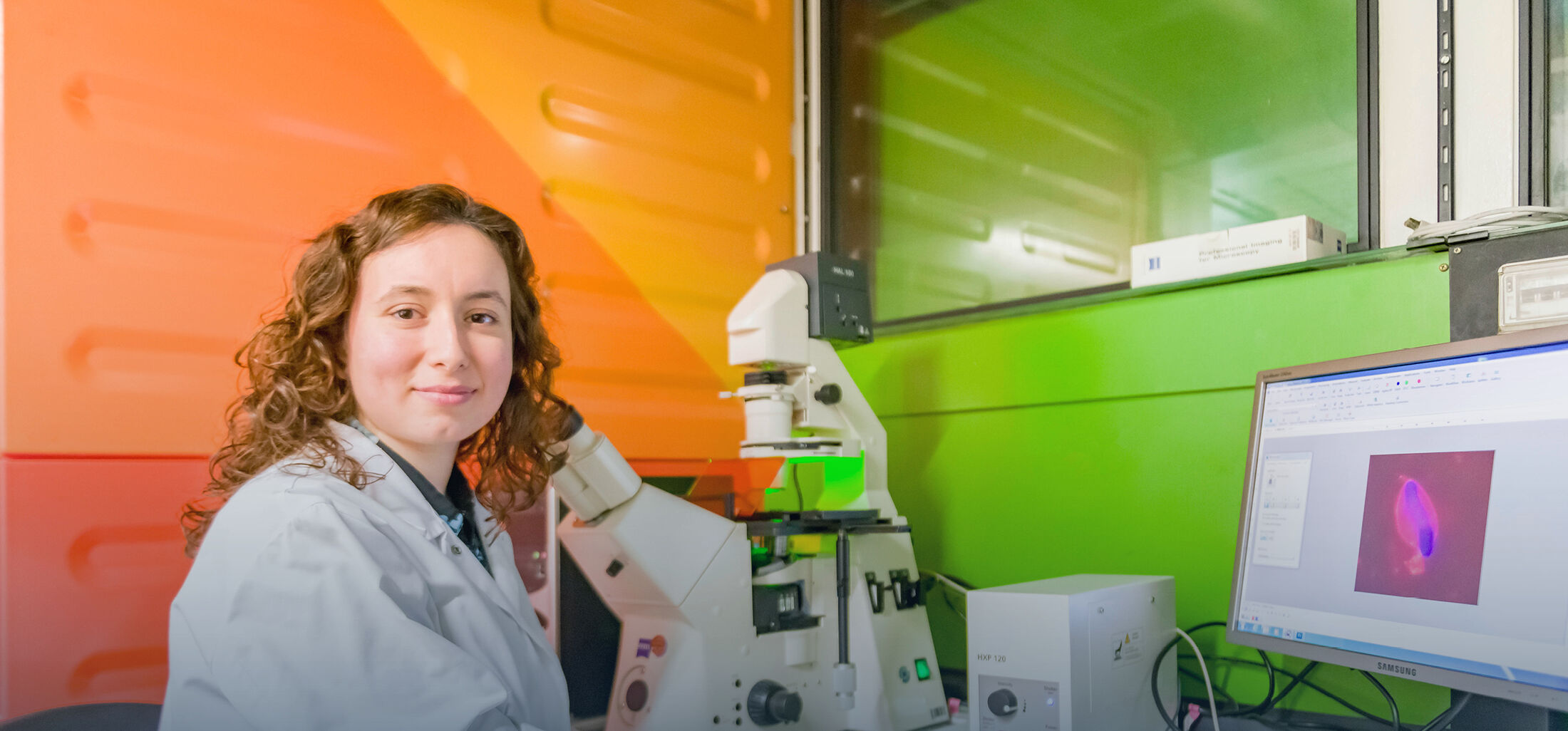
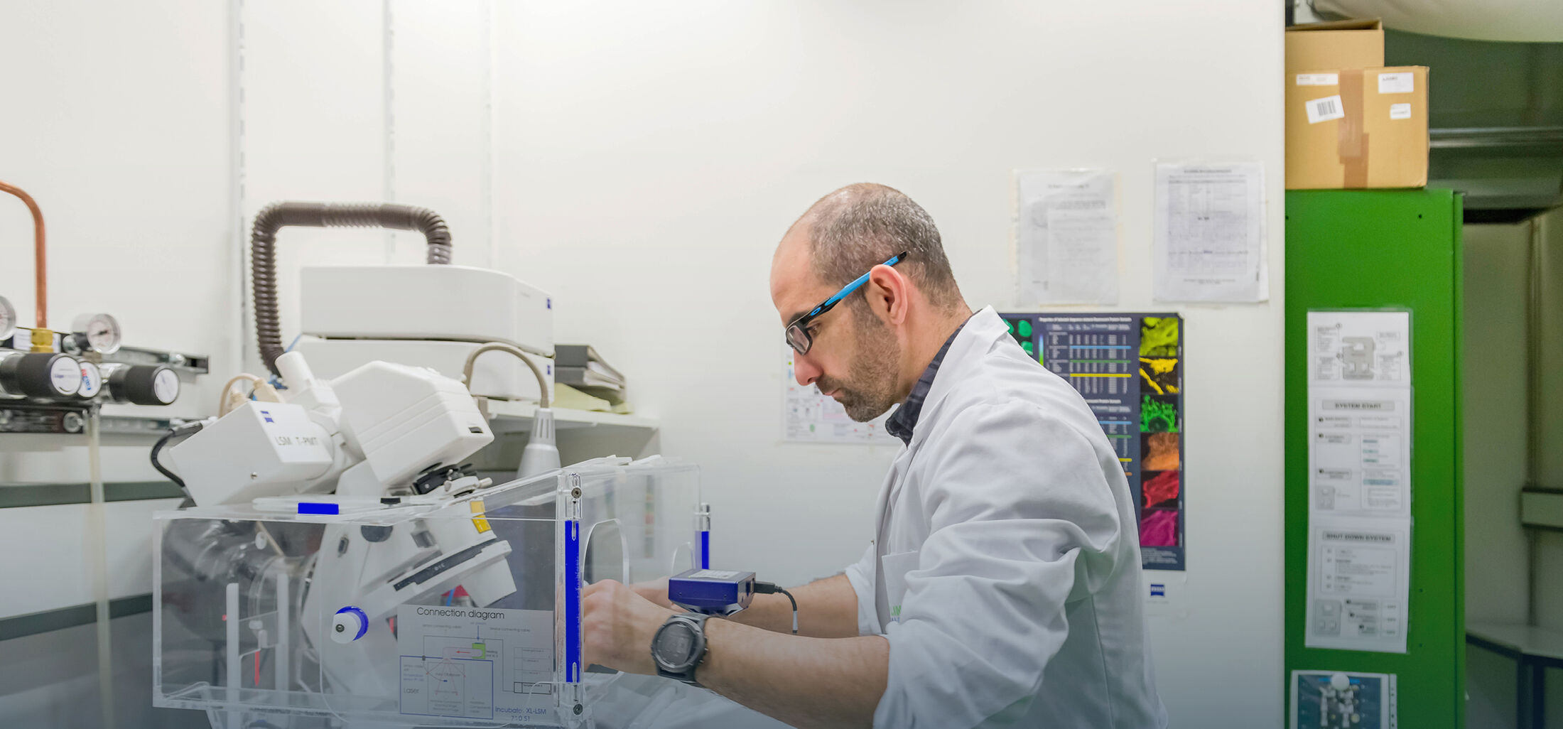
In the lab we use screening approaches using low- and high-resolution confocal microscopy to extract and analyze mitotic chromatin of live human cells. For this, we employ available state-of-the-art computational frameworks, but also continuously develop new algorithms to automatically segment and quantify mitotic chromatin.
Find out more about our research identifying eIF4A1/2 as the first RNA helicase regulating mitotic exit in this video.
Bachelor or master thesis project “chromatin decondensation at the end of mitosis”
We are looking for enthusiastic and motivated students (Biology, Biotechnology, Biochemistry, Chemistry, Biomedical Engineering or related disciplines) interested in a bachelor or master thesis project on chromatin decondensation at the end of mitosis at the Institute of Biochemistry and Molecular Cell Biology. The topic can be also addressed as research project for a medical thesis.
Mitosis is the most impressive stage of the cell cycle. During mitosis the chromatin structure changes dynamically: at the beginning of mitosis chromatin condenses into densely-packed structures to enable correct segregation of the genetic material; at the end of mitosis chromatin decondenses to re-establish the interphase chromatin structure, making it accessible for DNA replication and transcription. To understand the molecular mechanisms involved in chromatin decondensation at the end of mitosis, we use live-cell imaging of human cells. We develop techniques to generate and analyse low- and high-throughput imaging data, and draw hypotheses from these how specific chromatin decondensation factors are involved in this process.
Some research questions you could address in your project include:
- How is RNA as an architectural molecule on the periphery of mitotic chromosomes involved in chromatin decondensation?
- How can distinct RNA helicases by acting on the periphery of mitotic chromosomes influence the decondensation of chromatin?
You will use the following methods in the lab:
Cell culture, siRNA and DNA transfection, high resolution live cell imaging, western blot
Selected publications:
Magalska A, Schellhaus AK, Moreno-Andrés D, Zanini F, Schooley A, Sachdev R, Schwarz H, Madlung J, Antonin W (2014). RuvB-like ATPases Function in Chromatin Decondensation at the End of Mitosis. Dev Cell 31, 305–318.
Antonin W and Neumann H (2016). Chromosome condensation and decondensation during mitosis. Curr Opin Cell Biol 40, 15–22.
Moreno-Andrés D, Yokoyama H, Scheufen A, Holzer G, Lue H, Schellhaus AK, Weberruss M, Takagi M, Antonin W (2020). VPS72/YL1-Mediated H2A.Z Deposition Is Required for Nuclear Reassembly after Mitosis. Cells 9, E1702.
Moreno-Andrés D, Bhattacharyya A, Scheufen A, Stegmaier J (2022). LiveCellMiner: A new tool to analyze mitotic progression. PLoS One 17, e0270923.
Contact:
Dr. Ramona Jühlen
rjuehlenukaachende
Institute of Biochemistry and Molecular Cell Biology
Medical Faculty, RWTH Aachen University
Pauwelsstrasse 30
52074 Aachen