Epithelial-mesenchymal transition (EMT) in BCSCs
BCSCs show specific molecular profiles which likely account for their metastatic behavior including the organ tropism during tumor progression.
There is increasing evidence that an epithelial-to-mesenchymal transition (EMT) is a prerequisite to metastatic disease. Since CSCs are thought to be the motor behind tumor initiation, growth and eventually metastasis several groups have closely linked the existence of a CSC to EMT and showed that the metastatic cell, that underwent the EMT, is a CSC. We have developed a method to isolate and cultivate pure, highly tumorigenic CSCs from triple negative breast cancer from human patients (Metzger and Stepputtis et al, 2017). In addition, we could show that there are epithelial and mesenchymal CSCs. Some of the isolated CSC lines are also metastatic. Based on these findings we want to determine what kind of role the EMT plays in migratory/metastatic potential of primary human CSCs. To this end we will determine BCSC morphological and migratory behavior as well as their invasive, disseminating and metastatic capacity in patient derived xenograft (PDX) models. The underlying molecular mechanism explaining differences in these capacities will be investigated by unbiased screening (genomic and proteomic profiling) as well as candidate genes (EMT-, stemness-factors, such as transcription factors Slug, Zeb1, Vimentin, etc.) and pathway analyses. Here we could already help to identify new players like ESRP1 in the EMT paradigm (Preca et al., 2015, Preca et al., 2017) Newly identified markers are also validated for clinical relevance in human cancers and their prognostic value is determined. Ultimately we will use a screen for small molecular inhibitors to abolish EMT and stemness in CSCs in vitro and test these agents in vivo for their potential to inhibit metastasis.
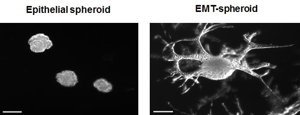
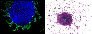
Upper panel: Morphology of epithelial and EMT-spheroids in 3D Matrix. Lower left panel: 2D culture of cancer stem cells with an epithelial core (DAPI, blue only) and cells on the edges that turn on Vimentin (green) and undergo an EMT. Lower right panel: Cancer stem cell culture undergoing EMT, stained with Crystal violet.


