Arbeitsgruppe MRT-Forschung
Die Arbeitsgruppe MRT-Forschung beschäftigt sich mit einer Vielzahl präklinischer und klinischer Fragestellungen:
- Techniken der MR-Bildakquise und -verarbeitung, auch unter Nutzung von Techniken der Künstlichen Intelligenz (Artificial Intelligence – AI)
- MR-Bildgebung der Leberfunktion
- Bildgebende Nachuntersuchungen nach interventionell-radiologischen Therapien
- Systematische Optimierung und Validierung neuartiger MR-Sequenzen, z.B. kombinierter morphologisch-funktioneller Sequenzen zur umfassenderen Gewebebeurteilung sowie neuer Techniken der Prostata-MRT
- Neue Methoden der MR-gesteuerten Prostatabiopsie
Team
Teresa is MRI physicist in the Department of Diagnostic and Interventional Radiology and works at the interface between physics and medicine. Her main research interest is quantitative MRI, i.e., to quantitatively characterize tissue parameters beyond the mere assessment of tissue morphology, but she also loves to work on and improve image acquisition and data post-processing, and the application of new technical developments within the clinical environment. Teresa performed her Ph.D. studies at the Institute for Experimental Molecular Imaging under supervision of Prof. Volkmar Schulz and at Philips Research Eindhoven between 2016 and 2020 and completed her Ph.D. thesis in physics (entitled “Magnetic resonance fingerprinting for quantitative imaging of the breast”) in 2021. Since the early days of her Ph.D., Teresa has a strong interest in interdisciplinary work. Her personal motivation is to foster the interaction between natural scientists and medical researchers to create a mutual benefit for both worlds.
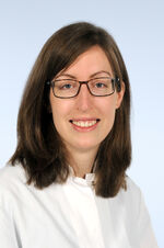
Alex is a senior clinical radiologist specialized in body imaging and imaging scientist with a special focus on liver magnetic resonance imaging and thrives to bring MR-based diagnosis, treatment planning, response assessment and prognostication to the next level by evaluating cutting edge MR-techniques in clinical practice. After graduating from RWTH Aachen University, she joined the Department of Diagnostic and Interventional Radiology (University Hospital Aachen) in 2010, where she started her research journey with her MD thesis on the comparison of PET/CT and liver MRI for response assessment after radioembolization. In 2015, she became deputy head of the department’s clinical MR-imaging section, just after receiving her board certification. Besides her clinical activities she pursued her research interests and co-founded the research group on liver MR-imaging. In 2019, she received a research grant from the medical faculty of RWTH Aachen University allowing her to expand her research activities. Since 2020 she is head of the clinical MR-imaging section at the Department of Diagnostic and Interventional Radiology.
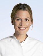
Elene joined the Department of Diagnostic and Interventional Radiology, as well as our team in 2023 and brought 5 years of research experience in MRI with her. She has worked in the research center Jülich on quantitative MRI and investigated its parameter changes in brain caused by several pathologies, including Parkinson’s disease, type 2 diabetes mellitus and obesity. In her Dr. med. Thesis (RWTH Aachen University), she characterized cerebral white matter hyperintensities using qMRI parameters and Fazekas scale to unveil the underlying pathology in vivo and noninvasively. She has also researched metabolic changes in the motor cortex as a result of transcranial direct current stimulation using GABA- and Phosphorus spectroscopy. In our team, Elene continues to pursue her strong interest in translational research and her fascination about complexity of the human body to improve the methods for a faster and more accurate diagnosis of various pathologies. She also improves her clinical skills in our department as she continuous her residency-training as a radiologist with hands-on experience.
Elisa is a medical student at RWTH Aachen University and she is currently working on her MD thesis in our team. Her research is focused on MRI-based, non-invasive quantification of fat in liver diseases. She is comparing the current standard, i.e., a breath hold sequence, to a free-breathing variant thereof. The goal is to eliminate breathing artifacts, especially in very young and in elderly patients.
Max is a medical student at Justus-Liebig-University in Gießen. He joined the team in 2023. Before he began studying medicine, he completed his training as a radiographer and is working since in the Department of Diagnostic and Interventional Radiology in Aachen, where he specified in the field of MR-Imaging. Joining his expertise and his interests, his PhD project investigates the influence of different respiratory triggering methods on MR image quality and workflow.
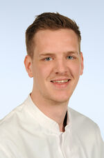
Sven is a clinical radiologist and imaging scientist with a focus on clinically motivated imaging research that aims to refine image acquisition and post-processing methodologies in close collaboration with clinicans, clinical scientists, engineers, MR physicists, and imaging scientists. After studying medicine at RWTH Aachen University, he completed his MD thesis on cartilage tissue engineering and joined the Department of Orthopedics (University Hospital Aachen) to receive orthopedic training during the surgical common trunk. After undertaking a period of research at the Institute of Anatomy (RWTH Aachen University) in 2015, he entered Radiology specialist training at the Department of Diagnostic and Interventional Radiology (University Hospital Aachen). After pursuing his research at the Department of Diagnostic and Interventional Radiology (University Hospital Düsseldorf) from 2019 to 2021, he moved back to Aachen to lead the group. His research is generously funded by the German Research Association (DFG) and by funds from RWTH AachenUniversity and Heinrich-Heine-University Düsseldorf. His recent publications are listed on Google Scholar and Pubmed. He also routinely reviews manuscripts for medical, technical, and interdisciplinary scientific journals.
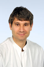
Shuo is Senior Clinical Scientist from Philips and Visiting Scholar at RWTH Aachen University Hospital (h-index 20). He has a university training background in Biomedical Engineering and holds a PhD degree in Medical Physics from the Max-Planck-Institute and Georg-August-University Göttingen, Germany. He is specialized in ultrafast imaging, cardiovascular imaging and quantitative imaging in the field of magnetic resonance (MR) technical development and new applications. He has worked as Research Scientist at the German Centre for Cardiovascular Research / Deutsches Zentrum für Herz-Kreislauf-Forschung (DZHK) and Philips Singapore for Asia Pacific. Since 2018 he has joined Philips Germany/DACH and Aachen's team in the Dept of Diagnostic and Interventional Radiology. His current interest as well as supervisory topics in MR research and development is breast care and pediatric care.
Aktuelle Publikationen:
T Lemainque: Editorial for "Effect of Phase Encoding Direction on Image Quality in Single-Shot EPI Diffusion-Weighted Imaging of the Breast". In: Journal of Magnetic Resonance Imaging
Teresa Lemainque, Marc Sebastian Huppertz, Can Yüksel, Robert Siepmann, Christiane Kuhl, Frank Roemer, Daniel Truhn, Sven Nebelung: Aktuelle MRT-Bildgebung des Knorpels im Kontext der Gonarthrose (Teil 1). In: Die Radiologie
Marc Sebastian Huppertz, Teresa Lemainque, Can Yüksel, Robert Siepmann, Christiane Kuhl, Frank Roemer, Daniel Truhn, Sven Nebelung: Aktuelle MRT-Bildgebung des Knorpels im Kontext der Gonarthrose (Teil 2). In: Die Radiologie
Ferhan Baskaya, Teresa Lemainque, Barbara Klinkhammer, Susanne Koletnik, Saskia von Stillfried, Steven Talbot, Peter Boor, Volkmar Schulz, Wiltrud Lederle, Fabian Kiessling: Pathophysiologic Mapping of Chronic Liver Diseases With Longitudinal Multiparametric MRI in Animal Models. In: Investigative Radiology
Gustav Müller-Franzes, Luisa Huck, Maike Bode, Sven Nebelung, Christiane Kuhl, Daniel Truhn, Teresa Lemainque: Diffusion probabilistic versus generative adversarial models to reduce contrast agent dose in breast MRI. In: European Radiology Experimental
Teresa Lemainque, Nicola Pridöhl, Shuo Zhang, Marc Huppertz, Manuel Post, Can Yüksel, Masami Yoneyama, Andreas Prescher, Christiane Kuhl, Daniel Truhn, Sven Nebelung: Time-efficient combined morphologic and quantitative joint MRI: an in situ study of standardized knee cartilage defects in human cadaveric specimens. In: European Radiology Experimental
Teresa Lemainque, Nicola Pridöhl, Marc Huppertz, Manuel Post, Can Yüksel, Robert Siepmann, Karl Ludger Radke, Shuo Zhang, Masami Yoneyama, Andreas Prescher, Christiane Kuhl, Daniel Truhn, Sven Nebelung: Two for One—Combined Morphologic and Quantitative Knee Joint MRI Using a Versatile Turbo Spin-Echo Platform. In: Diagnostics
Teresa Lemainque, Masami Yoneyama, Chiara Morsch, Elene Iordanishvili, Alexandra Barabasch, Maximilian Schulze-Hagen, Johannes M Peeters, Christiane Kuhl, Shuo Zhang: Reduction of ADC bias in diffusion MRI with deep learning-based acceleration: A phantom validation study at 3.0 T. In: Magnetic Resonance Imaging
Sarah Schraven, Ramona Brück, Stefanie Rosenhain, Teresa Lemainque, David Heines, Hormoz Noormohammadian, Oliver Pabst, Wiltrud Lederle, Felix Gremse, Fabian Kiessling. CT-and MRI-Aided Fluorescence Tomography Reconstructions for Biodistribution Analysis. In: Investigative Radiology.
Müller-Franzes G, Niehues JM, Khader F, Arasteh ST, Haarburger C, Kuhl C, Wang T, Han T, Nolte T, Nebelung S, Kather JN, Truhn D
A multimodal comparison of latent denoising diffusion probabilistic models and generative adversarial networks for medical image synthesis.
In: Scientific reports
Thomas A, Nolte T, Baragona M, Ritter A
Finding an effective MRI sequence to visualise the electroporated area in plant-based models by quantitative mapping.
In: Bioelectrochemistry (Amsterdam, Netherlands)
Müller-Franzes G, Huck L, Tayebi Arasteh S, Khader F, Han T, Schulz V, Dethlefsen E, Kather JN, Nebelung S, Nolte T, Kuhl C, Truhn D
Using Machine Learning to Reduce the Need for Contrast Agents in Breast MRI through Synthetic Images.
In: Radiology
Lindelauf KHK, Thomas A, Baragona M, Jouni A, Nolte T, Pedersoli F, Pfeffer J, Baumann M, Maessen RTH, Ritter A
Plant-based model for the visual evaluation of electroporated area after irreversible electroporation and its comparison to in-vivo animal data.
In: Science progress
Bode M, Charlotte Huck L, Zhang S, Nolte T, Yoneyama M, Nebelung S, Katharina Kuhl C
Clinical evaluation of cylindrical regional suppression in dynamic contrast-enhanced breast MRI: An intra-individual comparison study on image quality and lesion conspicuity.
In: European journal of radiology
Nolte T, Westfechtel S, Schock J, Knobe M, Pastor T, Pfaehler E, Kuhl C, Truhn D, Nebelung S
Getting Cartilage Thickness Measurements Right: A Systematic Inter-Method Comparison Using MRI Data from the Osteoarthritis Initiative.
In: Cartilage
Zanderigo E, Huck L, Distelmaier M, Dethlefsen E, Maywald M, Truhn D, Dirrichs T, Doneva M, Schulz V, Kuhl CK, Nolte T
Feasibility study of 2D Dixon-Magnetic Resonance Fingerprinting (MRF) of breast cancer.
In: European journal of radiology open
Bode M, Zhang S, Terwolbeck MN, Tabrizi CM, Sprenger P, Yoneyama M, Kraemer NA, Kuhl CK, Barabasch A
Author Correction: Liver diffusion‑weighted MR imaging with L1‑regularized iterative sensitivity encoding reconstruction based on single‑shot echo‑planar imaging: initial clinical experience.
In: Scientific reports
Bode M, Zhang S, Terwolbeck MN, Molavi Tabrizi C, Sprenger P, Yoneyama M, Kraemer NA, Kuhl CK, Barabasch A
Liver diffusion-weighted MR imaging with L1-regularized iterative sensitivity encoding reconstruction based on single-shot echo-planar imaging: initial clinical experience.
In: Scientific reports
Ciba M, Winkelmeyer EM, Schock J, Westfechtel S, Nolte T, Knobe M, Prescher A, Kuhl C, Truhn D, Nebelung S
Varus stress MRI in the refined assessment of the posterolateral corner of the knee joint.
In: Scientific reports
Pathak V, Roemhild K, Schipper S, Groß-Weege N, Nolte T, Ruetten S, Buhl EM, El Shafei A, Weiler M, Martin L, Marx G, Schulz V, Kiessling F, Lammers T, Koczera P
Theranostic Trigger-Responsive Carbon Monoxide-Generating Microbubbles.
In: Small (Weinheim an der Bergstrasse, Germany)
Müller-Franzes G, Nolte T, Ciba M, Schock J, Khader F, Prescher A, Wilms LM, Kuhl C, Nebelung S, Truhn D
Fast, Accurate, and Robust T2 Mapping of Articular Cartilage by Neural Networks.
In: Diagnostics (Basel, Switzerland)
Rama E, Mohapatra SR, Melcher C, Nolte T, Dadfar SM, Brueck R, Pathak V, Rix A, Gries T, Schulz V, Lammers T, Apel C, Jockenhoevel S, Kiessling F
Monitoring the Remodeling of Biohybrid Tissue-Engineered Vascular Grafts by Multimodal Molecular Imaging.
In: Advanced science (Weinheim, Baden-Wurttemberg, Germany)
Hitpass L, Distelmaier M, Neumann UP, Schöning W, Isfort P, Keil S, Kuhl CK, Bruners P, Barabasch A
Recurrent Colorectal Liver Metastases in the Liver Remnant After Major Liver Surgery-IRE as a Salvage Local Treatment When Resection and Thermal Ablation are Unsuitable.
In: Cardiovascular and interventional radiology
Said O, Schock J, Abrar DB, Schad P, Kuhl C, Nolte T, Knobe M, Prescher A, Truhn D, Nebelung S
In-Situ Cartilage Functionality Assessment Based on Advanced MRI Techniques and Precise Compartmental Knee Joint Loading through Varus and Valgus Stress.
In: Diagnostics (Basel, Switzerland)
Ciba M, Winkelmeyer EM, Schock J, Schad P, Kotowski N, Nolte T, Wollschläger LM, Knobe M, Prescher A, Kuhl C, Truhn D, Nebelung S
Comprehensive Assessment of Medial Knee Joint Instability by Valgus Stress MRI.
In: Diagnostics (Basel, Switzerland)
Nolte T, Vaidya N, Baragona M, Elevelt A, Lavezzo V, Maessen R, Schulz V, Veroy K
Study of flow effects on temperature-controlled radiofrequency ablation using phantom experiments and forward simulations.
In: Medical physics
Pathak V, Nolte T, Rama E, Rix A, Dadfar SM, Paefgen V, Banala S, Buhl EM, Weiler M, Schulz V, Lammers T, Kiessling F
Molecular magnetic resonance imaging of Alpha-v-Beta-3 integrin expression in tumors with ultrasound microbubbles.
In: Biomaterials
Hitpass L, Amygdalos I, Sieben P, Raaff V, Lang S, Bruners P, Kuhl CK, Barabasch A
MRI-Based Quantitation of Hepatic Steatosis Does Not Predict Hypertrophy Rate after Portal Vein Embolization in Patients with Colorectal Liver Metastasis and Normal to Moderately Elevated Fat Fraction.
In: Journal of clinical medicine
Nolte T, Scholten H, Gross-Weege N, Amthor T, Koken P, Doneva M, Schulz V
Confounding factors in breast magnetic resonance fingerprinting: B 1 + , slice profile, and diffusion effects.
In: Magnetic resonance in medicine
Bednarsch J, Czigany Z, Lurje I, Trautwein C, Lüdde T, Strnad P, Gaisa NT, Barabasch A, Bruners P, Ulmer T, Lang SA, Neumann UP, Lurje G
Intraoperative Transfusion of Fresh Frozen Plasma Predicts Morbidity Following Partial Liver Resection for Hepatocellular Carcinoma.
In: Journal of gastrointestinal surgery : official journal of the Society for Surgery of the Alimentary Tract
- Ruarus AH, Barabasch A, Catalano O, Leen E, Narayanan G, Nilsson A, Padia SA, Wiggermann P, Scheffer HJ, Meijerink MR
Irreversible Electroporation for Hepatic Tumors: Protocol Standardization Using the Modified Delphi Technique.
In: Journal of vascular and interventional radiology : JVIR. - Nolte T, Gross-Weege N, Schulz V
(Hybrid) SPECT and PET Technologies.
In: Recent results in cancer research. Fortschritte der Krebsforschung. Progres dans les recherches sur le cancer. - Theek B, Nolte T, Pantke D, Schrank F, Gremse F, Schulz V, Kiessling F
Emerging methods in radiology.
In: Der Radiologe. - Thüring J, Kuhl CK, Barabasch A, Hitpass L, Bode M, Bünting N, Bruners P, Krämer NA
Signal changes in T2-weighted MRI of liver metastases under bevacizumab-A practical imaging biomarker?
In: PloS one. - Nolte T, Gross-Weege N, Doneva M, Koken P, Elevelt A, Truhn D, Kuhl C, Schulz V
Spiral blurring correction with water-fat separation for magnetic resonance fingerprinting in the breast.
In: Magnetic resonance in medicine.
- Barabasch A, von Stillfried S, Kuhl CK, Heinzel A, Sander A, Isfort P
Toward Transpulmonary Chemoembolization with Degradable Starch Microspheres: Systematic Analysis of Local and Systemic Effects in a Porcine Model.
In: Cardiovascular and interventional radiology. - Gross-Weege N, Nolte T, Schulz V
MR image corrections for PET-induced gradient distortions.
In: Physics in medicine and biology. - Truhn D, Kuhl CK, Ciritsis A, Barabasch A, Kraemer NA
A New Model for MR Evaluation of Liver Function with Gadoxetic Acid, Including Both Uptake and Excretion.
In: European radiology.
- Ritter A, Bruners P, Isfort P, Barabasch A, Pfeffer J, Schmitz J, Pedersoli F, Baumann M
Electroporation of the Liver: More Than 2 Concurrently Active, Curved Electrodes Allow New Concepts for Irreversible Electroporation and Electrochemotherapy.
In: Technology in cancer research & treatment. - Barabasch A, Heinzel A, Bruners P, Kraemer NA, Kuhl CK
Diffusion-weighted MRI Is Superior to PET/CT in Predicting Survival of Patients Undergoing 90Y Radioembolization of Hepatic Metastases.
In: Radiology.
- istelmaier M, Barabasch A, Heil P, Kraemer NA, Isfort P, Keil S, Kuhl CK, Bruners P
Midterm Safety and Efficacy of Irreversible Electroporation of Malignant Liver Tumors Located Close to Major Portal or Hepatic Veins.
In: Radiology. - Barabasch A, Distelmaier M, Heil P, Krämer NA, Kuhl CK, Bruners P
Magnetic Resonance Imaging Findings After Percutaneous Irreversible Electroporation of Liver Metastases: A Systematic Longitudinal Study.
In: Investigative radiology.
- Barabasch A, Kraemer NA, Ciritsis A, Hansen NL, Lierfeld M, Heinzel A, Trautwein C, Neumann U, Kuhl CK
Diagnostic accuracy of diffusion-weighted magnetic resonance imaging versus positron emission tomography/computed tomography for early response assessment of liver metastases to Y90-radioembolization.
In: Investigative radiology. - Strobel K, Schrading S, Hansen NL, Barabasch A, Kuhl CK
Assessment of BI-RADS category 4 lesions detected with screening mammography and screening US: utility of MR imaging.
In: Radiology.
Bei Fragen bezüglich der Forschungsprojekte oder Interesse an aktiver Mitarbeit im Rahmen von Abschluss- oder Doktorarbeiten können Sie uns jederzeit gerne kontaktieren:
Dr. rer. nat. Teresa Lemainque (Teamleiterin)
tlemainqueukaachende
Dr. med. Alexandra Barabasch
abarabasch@ukaachen.de
PD Dr. med. Sven Nebelung
snebelungukaachende

