
Differentiation therapy targeting breast cancer stem cells
Our lab is working on the basis of the cancer stem cell (CSC) hypothesis: There is a certain amount of cancer stem cells in a given tumor which drive its progression and growth. The majority of the cells in a tumor would be differentiated progeny of said cells and would not contribute to the major problem of tumor progression, metastasis. One problem, to date, is the limited propagation of primary undifferentiated breast cancer stem cells in culture. This has impacted strategies of high throughput screening for therapeutics that might exploit the differentiation potential of CSC to control tumor progression and metastasis. Since we solved this problem of isolating of BCSSs (Castro and Maurer et al. 2013, Metzger and Stepputtis et al, 2017) we can now focus on several promising strategies to target these cells.
We are focusing here on differentiation therapy to potentially treat tumors which are driven by cancer stem cells. The idea is that whatever cell type was the origin of the CSC, it should be possible to “push” the CSC back into the normal process of differentiation. Upon entering a differentiation program, a CSC would stop being a tumor driver and become harmless tissue. A first success in this area could already be achieved using kinase knockdowns of ALPK1 and ERN1 in an attempt a bipotent triple negative breast cancer cell (Strietz et al., 2016). The fact that this approach does not involve directly killing cells, might make a therapy much less harmful for the patient, compared to chemotherapy. Our lab utilizes screening methods and biochemical experiments to identify agents that tip the scale from uncontrolled stem cell proliferation to differentiation. As basis for these experiments we use primary patient CSCs to address cancer as close to the patient as possible.
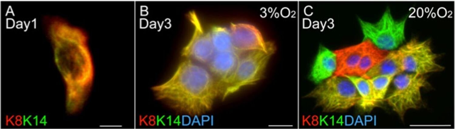
From left to right: cancer stem cells in 2D culture, single cells on day 1 in hypoxia, clonal cell colonies on day 3 in hypoxia (left) and normoxia (right). Already after 3 days differentiation of cancer stem cells can occur in normoxia. Immunohistochemistry of Keratin 8 (red), Keratin 14 (green) and DAPI (blue). Luminal (red) and myoepithelial (green) differentiation is seen after three days in normoxia in contrast to bi-potent cancer stem cells (yellow overlay).