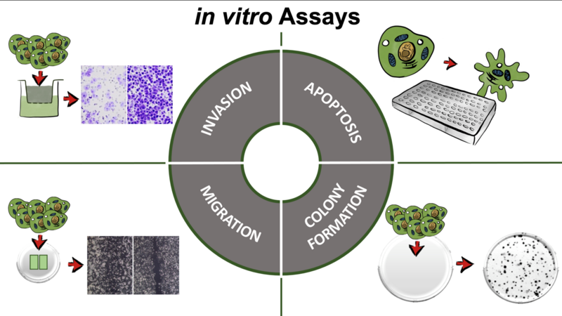
In vitro Assays
We are using a variety of functional in vitro assays to measure different tumor-cell specific characteristics in cell culture. For example, to test the ability of tumor cells to invade and migrate, a chemotactic gradient can be used. In addition, determining how long cells need to close a preformed gap the capacity to migrate can be assessed. In an apoptosis assay, cell death is induced to determine basal and induced apoptosis. Here, cell death is measured by the amount of resulting fluorescent product. In a colony formation assay a low number of cells is seeded and observed over a period of time. It may provide details about a cells capacity to form colonies, the colony morphology and the interactions of the cells involved.
Stable cell transfection
Class II tumor suppressor genes (C2TSGs) are frequently silenced in cancer cells by DNA promoter hypermethylation. To analyze the function of novel putative C2TSGs, cancer cell lines stably expressing the candidate gene are established. This is done by transfection using a vector containing the gene of interest and the genetic information for an antibiotic resistance. As a result, only successfully transfected cells will survive under selection pressure and start forming colonies after very thin cell seeding. After growing back these clones to considerable numbers, RT-PCR and Western Blot are performed to confirm successful integration and expression on mRNA and protein level. Generated stable clones containing either the gene of interest or empty vector (as control) are then subjected to various in vitro assays (see there) to learn more about C2TSG function in suppressing cancer.
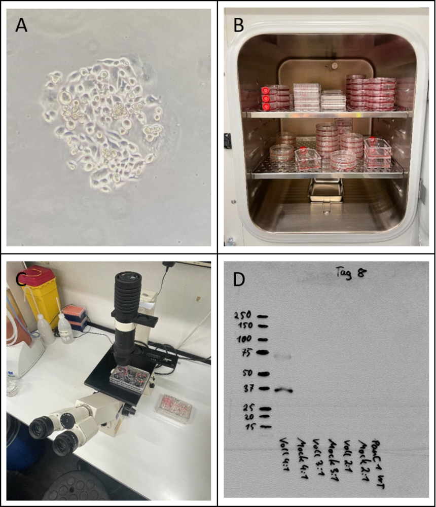
A) A forming colony of PANC1 cells, a pancreatic cancer cell line; B) Growing back from single cell clones up to the amount of whole petri dishes requires different sizes of cultivation wells. Different dish sizes can be found in this cell culture incubator; C) Transfected cells are getting monitored under the microscope regularly to observe their confluence; D) Typical western blot used to demonstrate protein expression of the gene of interest in transfected cells.
Tissue microarrays
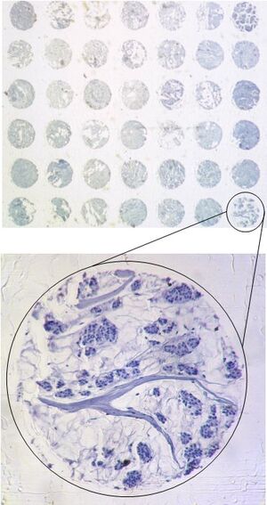
Similar to gene analysis, the trend in tissue analysis is towards high-throughput procedures. According to a method originally established by Kallioniemi & Sauter (Kononen et al. 1998: Tissue microarrays for high-throughput molecular profiling of tumor specimens. Nat Med. 1998 4:844-7) up to several hundred different tissue samples (e. g. tumors) are arrayed on a single glass slide. As with the DNA array, each position of the array is clearly characterized by its coordinates.
Tissue Micro Array technology allows the simultaneous analysis of many tissue samples. This saves time and money (e. g. for antibodies). The spot size of the tissues on the array is usually between 0.6 and 1.2 mm. You can see an excerpt from a breast cancer tissue micro array below.