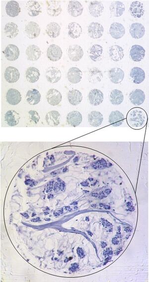


Tissue microarrays
Similar to gene analysis, the trend in tissue analysis is towards high-throughput procedures. According to a method originally established by Kallioniemi & Sauter (Kononen et al. 1998: Tissue microarrays for high-throughput molecular profiling of tumor specimens. Nat Med. 1998 4:844-7) up to several hundred different tissue samples (e. g. tumors) are arrayed on a single glass slide. As with the DNA array, each position of the array is clearly characterized by its coordinates.
Tissue Micro Array technology allows the simultaneous analysis of many tissue samples. This saves time and money (e. g. for antibodies). The spot size of the tissues on the array is usually between 0.6 and 1.2 mm. You can see an excerpt from a breast cancer tissue micro array below.