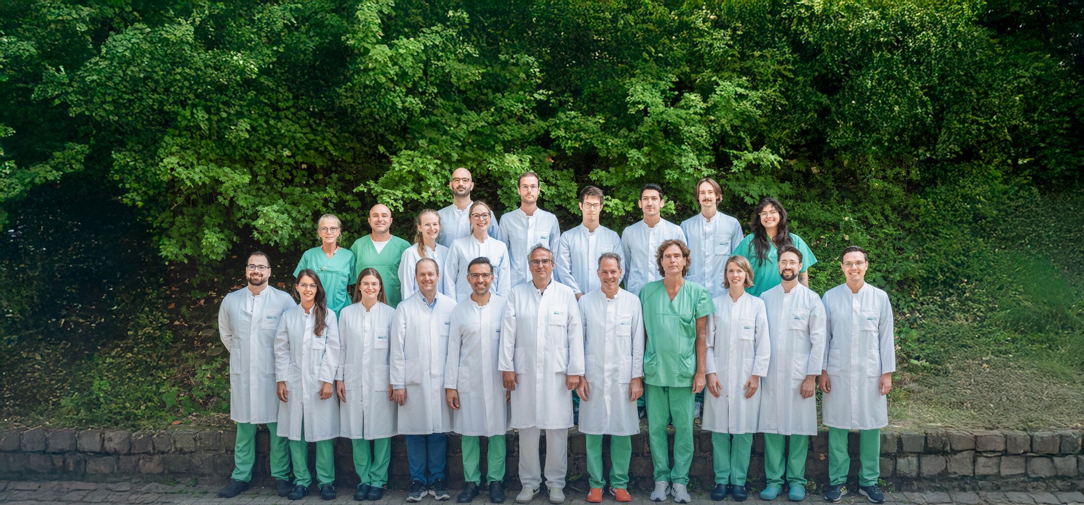

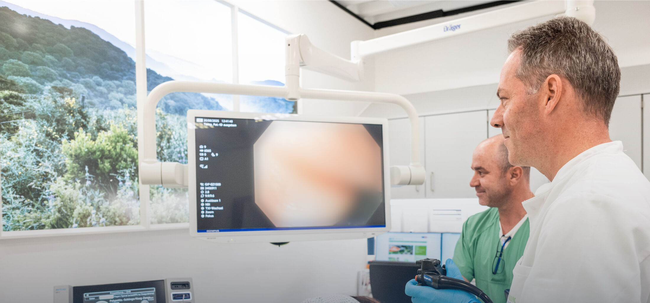
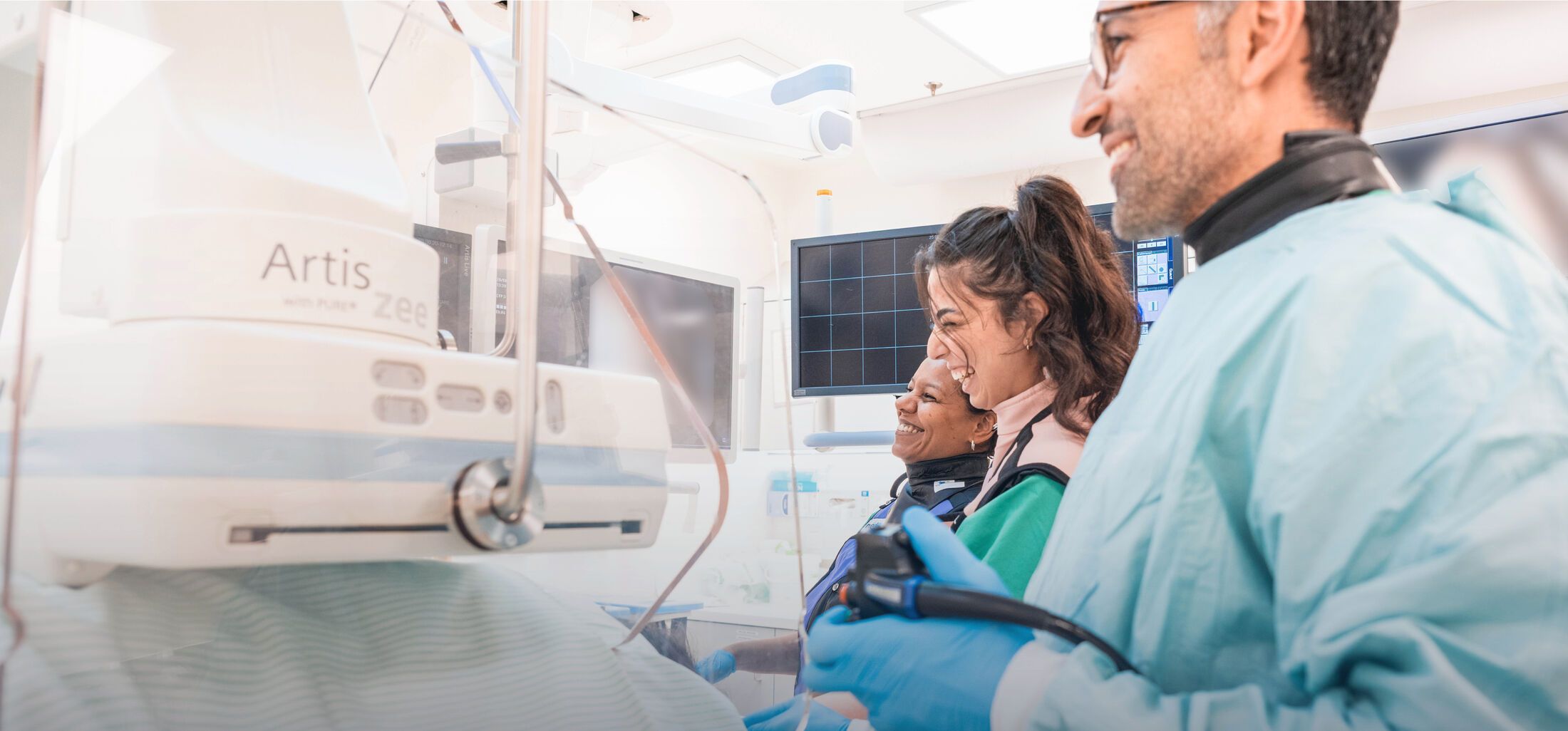
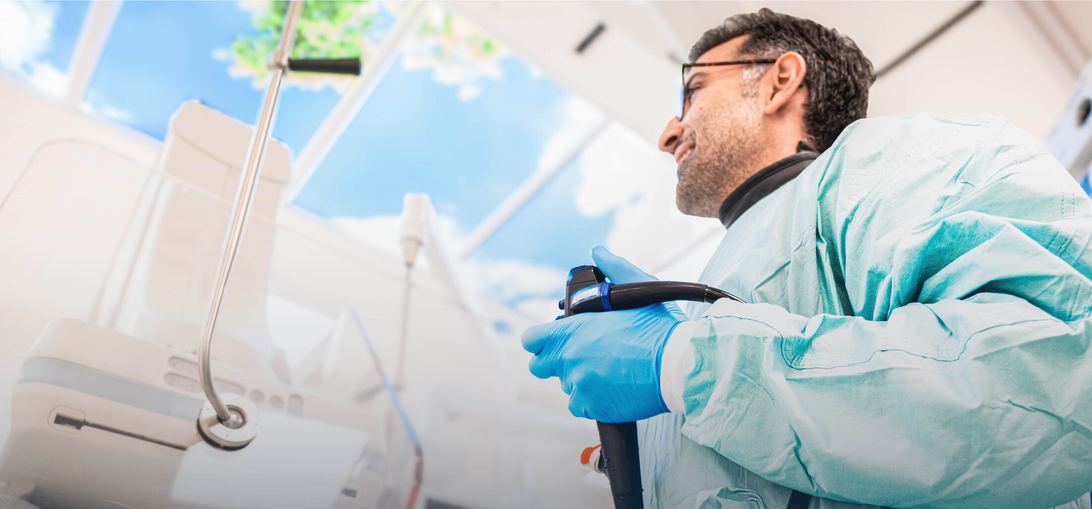
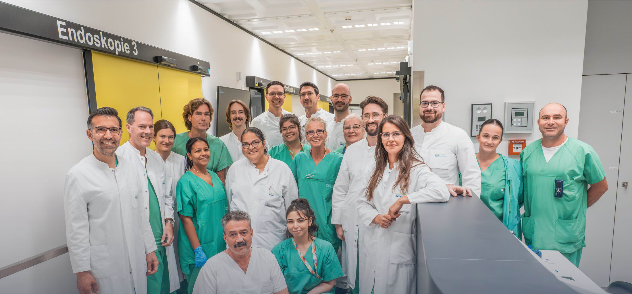

In addition to first-class equipment with the newest high-end devices and nearly all disposable instruments from various manufacturers, our interdisciplinary endoscopy unit has several experienced and certified physicians. Our physicians work closely together on an interdisciplinary basis in a central unit, undertake regular continuing education, and train physicians from other clinics, both nationally and internationally. We are therefore able to offer you in the field of endoscopy all standard as well as innovative procedures at the highest quality standard. As a university hospital (so-called "supramaximal care"), we furthermore have the ability to coordinate quickly and individually with all medical specialties.
We would like to briefly introduce, in plain and understandable language, the principal procedures that we offer as a matter of routine. During an outpatient or inpatient stay, we will of course be glad to explain to you the background of the procedures that may be relevant to you, and the advantages and disadvantages that these have for your individual situation. The many innovative procedures, which are constantly being further developed, cannot be presented here in full; we will be glad to explain these to you in a personal consent discussion tailored to your individual situation.
Endoscopy of the upper gastrointestinal tract (esophagogastroduodenoscopy (EGD), gastroscopy, or, colloquially, "gastroscopy"/"stomach endoscopy") serves the diagnosis and treatment of diseases and changes in the upper gastrointestinal tract (esophagus, stomach, and duodenum). Samples can be taken during the examination, and special techniques or staining methods (virtual or dye-assisted chromoendoscopy) can be used to detect early changes before a malignant disease develops. Many treatment options exist for therapy, some of which can also be carried out on an outpatient basis, for example:
- Removal of tumors (advanced polyps or early cancers, so-called "early carcinomas") via the endoscope, without any incision from outside
- Removal of masses (subepithelial lesions) via the endoscope, without any incision from outside
- Resolution of swallowing disorders (e.g. by means of peroral endoscopic myotomy (POEM), balloon dilation, or Botox injection)
- Widening of strictures (stenoses) by means of dilation (bougienage or dilation) or the insertion of stents
- Treatment of bleeding or vascular changes (all techniques)
- Surveillance / dysplasia screening for Barrett's esophagus or tumors in the ENT region
The following technical options are available:
- High-resolution esophagogastroduodenoscopy (EGD)
- Zoom endoscopy (optical and digital)
- Chromoendoscopy and virtual chromoendoscopy
- Application of various imaging technologies:
- NBI (Narrow Band Imaging)
- TXI (Texture and Color Enhancement Imaging)
- RDI (Red Dichromatic Imaging)
- LCI (Linked Color Imaging)
- BLI (Blue Light Imaging)
- ACI (Amber-Red Colour Imaging)
- Endoscopes of the widest range of diameters and lengths from two leading endoscope manufacturers (Olympus and Fujifilm) for full-scope treatment (e.g. removal of early carcinomas or POEM for motility disorders)
Using longer endoscopes and a special endoscopic technique employing a balloon around the endoscope, deeper parts of the small bowel can be examined, either from the oral route (from the mouth) or from the anal route (from the anus), and treated directly where necessary. The small bowel can also be visualized using a video capsule, which contains a small camera and is swallowed. These techniques are used in particular when searching for possible sources of bleeding, tumors, or vascular malformations (angiodysplasias), strictures, or inflamed areas.
The following technical options are available:
- Capsule endoscopy
- Push enteroscopy
- Balloon enteroscopy (single-balloon (SBE) and double-balloon (DBE))
- Balloon-ERCP (in altered anatomy, a combination of balloon enteroscopy and procedures on the bile ducts or pancreatic duct)
In endoscopy of the lower gastrointestinal tract, the large bowel (colon) and, where relevant, the last section of the small bowel (terminal ileum) are examined. Beyond colorectal cancer screening with removal of polyps, tumors can also be removed endoscopically, and strictures (stenoses) can be widened or treated with stents. New developments now allow us, during the examination in several of our rooms, to additionally employ artificial intelligence to support the detection and assessment of polyps.
Samples can be taken during the examination, and special techniques or staining methods (virtual or dye-assisted chromoendoscopy) can be used to detect early changes before a malignant disease develops. Many treatment options exist for therapy, some of which can also be carried out on an outpatient basis, for example:
- Removal of tumors (advanced polyps or early cancers, so-called "early carcinomas") via the endoscope, without any incision from outside
- Removal of masses (subepithelial lesions) via the endoscope, without any incision from outside
- Widening of strictures (stenoses) by means of dilation (bougienage or dilation) or the insertion of stents
- Treatment of bleeding or vascular changes (all techniques)
- Surveillance / dysplasia screening for chronic inflammatory bowel disease (IBD)
The following technical options are available:
- High-resolution ileocolonoscopy/sigmoidoscopy
- Chromoendoscopy and virtual chromoendoscopy
- Zoom endoscopy (optical and virtual)
- Rectoscopy
- Proctoscopy
- Polyp detection using artificial intelligence (Olympus and Fujifilm)
- Polyp measurement using AI-supported laser technology
- Application of various imaging technologies:
- NBI (Narrow Band Imaging)
- TXI (Texture and Color Enhancement Imaging)
- RDI (Red Dichromatic Imaging)
- LCI (Linked Color Imaging)
- BLI (Blue Light Imaging)
- ACI (Amber-Red Colour Imaging)
- Endoscopes of the widest range of diameters and lengths from two leading endoscope manufacturers (Olympus and Fujifilm) for full-scope treatment (e.g. removal of polyps and early tumors, or treatment of strictures)
In our department, an examination of the esophageal musculature is possible for patients with swallowing difficulties (dysphagia). Using high-resolution esophageal manometry (HR manometry) or impedance planimetry (the so-called "EndoFLIP" technique), the swallowing act can be examined and classified. In addition, the reflux of stomach acid into the esophagus (reflux) can be examined by means of 24-hour pH-metry (where relevant, together with impedance testing). For this, a probe is inserted via the nose, and the measurement result is evaluated the following day.
We also offer breath tests for the investigation of various conditions. For this, following appropriate preparation over the preceding days, a corresponding drinking solution is consumed. Metabolic processes in the bowel produce hydrogen (H2), which can be measured in exhaled breath and can provide indications of dysfunctions or bacterial overgrowth (SIBO).
The following technical options are available:
- High-resolution esophageal and rectal manometry (HR manometry)
- Impedance planimetry of the esophagus, the pylorus, or the anal sphincter (using the EndoFLIP system)
- 24-hour pH-metry and impedance measurement
- H2 breath tests (lactose, fructose, glucose, lactulose, xylose)
- Rapid lactase test from duodenal biopsies
Further information can be found here.
In our modern endoscopy unit, the entire spectrum of endoscopic diagnostics and therapy is possible. Suspicious changes and tumors can be removed from the mucosa in various ways, which may spare you major surgical procedures. In addition, mucosal changes can be treated with argon plasma (APC) or heat by means of radiofrequency ablation (RFA), for example to prevent tumor formation or bleeding. When it is no longer possible to take in food, or when bile or pancreatic fluid can no longer drain properly, various methods are available to us to improve this. Varicose veins in the esophagus or hemorrhoids in the rectum can be tied off using rubber bands (ligation). For disorders of the musculature, a Botox injection, dilation (balloon dilation), or splitting of the musculature (myotomy, e.g. by peroral endoscopic myotomy (POEM)) can be carried out.
Therapeutic spectrum:
- Removal/resection of polyps or early tumors/cancers (so-called "early carcinomas") in all gastrointestinal organs (e.g. endoscopic submucosal dissection (ESD), endoscopic intermuscular dissection (EID), or full-thickness resection (full-thickness resection device (fTRD) or knife-assisted full-thickness resection (kFTR)))
- Removal/resection of masses (subepithelial lesions) (e.g. endoscopic submucosal dissection (ESD), endoscopic intermuscular dissection (EID), tunnelling techniques (e.g. submucosal tunnelling and endoscopic resection (STER)), or full-thickness resection (full-thickness resection device (fTRD) or knife-assisted full-thickness resection (kFTR)))
- Peroral endoscopic myotomy (POEM, e.g. esophagus and stomach)
- Splitting for Zenker's diverticulum (POEM or mucomyotomy)
- Endoscopic closure techniques (endoscopic suturing (e.g. SutuArt, X-Tack), endoscopic vacuum therapy, various clips)
- Bougienage of the esophagus or rectum
- Balloon dilation in the esophagus, stomach, or small/large bowel
- Botox injections in the esophagus, stomach, or small/large bowel
- Stent insertion, exchange, and removal in the esophagus, stomach, small/large bowel, bile ducts, or pancreatic duct
- Ablation procedures (e.g. radiofrequency ablation (RFA), argon plasma coagulation (APC))
- Hemostasis techniques (all standard methods)
- Endoluminal vacuum therapy (with sponge or stent)
- PEG tube placement, exchange, and removal
- Endosonographic punctures (fine-needle aspiration (FNA) or fine-needle biopsy (FNB); e.g. for the investigation of a mass)
- Endosonographic drainages (all techniques)
- Endosonographic anastomoses (e.g. gastroenterostomy (GE), hepaticogastrostomy (HGS), choledochoduodenostomy (CDS), pancreatic duct drainage)
- Procedures on the bile ducts via ERCP, balloon-ERCP, endosonography, or PTCD (combined where appropriate)
- Procedures on the pancreatic duct via ERCP, endosonography, or balloon-ERCP (combined where appropriate)
- Endoscopic tumor therapy for non-removable tumors (e.g. RFA in the bile duct)
- Endoscopic reflux therapy (e.g. anti-reflux mucosal ablation (ARMA) or anti-reflux mucosectomy (ARMS))
- Bariatric procedures
If, as a colleague, you have questions on this topic or would like to refer your patients to us, we can gladly plan corresponding procedures promptly in our Intervention Consultation.
As with a conventional gastroscopy or colonoscopy, special endoscopes fitted with an ultrasound head can be introduced via the mouth or the anus. This allows an ultrasound examination from the inside (endoluminal), and offers a better assessment for many conditions than a conventional ultrasound from the outside (transcutaneous), as well as a higher resolution than other imaging procedures (e.g. CT). In addition, under endoscopic ultrasound guidance, fluid collections can be punctured and samples taken from, for example, tumors. The diversion of bile or pancreatic fluid into the gastrointestinal tract is also possible through the insertion of special stents. Furthermore, using metal stents, a connection can be created between, for example, the stomach and bowel (gastroenterostomy) or the stomach and liver (hepaticogastrostomy). In the case of pancreatitis or postoperatively, collections or areas of necrosis can be diverted into the stomach or bowel.
Diagnostic spectrum:
- Longitudinal and radial endosonography from two renowned manufacturers (Olympus and Fujifilm)
- Contrast-enhanced endosonography
- Elastography
- Fine-needle biopsy and aspiration (FNB and FNA)
Therapeutic spectrum:
- Drainage placement for peripancreatic fluid collections (e.g. for pancreatitis or postoperatively)
- Diversion of the bile ducts into the gastrointestinal tract (endosonographic bilio-digestive anastomoses, e.g. hepaticogastrostomy (HGS), choledochoduodenostomy (CDS), or antegrade stenting)
- Diversion of pancreatic fluid into the gastrointestinal tract (endosonographic pancreatic duct drainage)
- Connection between stomach and bowel (endosonographic gastroenterostomy)
- Procedures on the bile ducts or pancreas in altered anatomy (e.g. connection of the remnant stomach and bypassed stomach in gastric bypass patients using EDGE (endosonography-guided transgastric ERCP), or connection of small bowel segments using EDEE (endosonography-guided transenteric ERCP))
- Therapy for chronic abdominal pain (ganglion block)
- Necrosectomy (endoscopic, or combined endoscopic and percutaneous)
An endoscopic retrograde cholangiopancreatography (ERCP) visualizes the bile ducts of the liver and/or the pancreas. The examination is usually carried out with the patient lying prone. As with a gastroscopy, the endoscope is advanced as far as the small bowel, and the bile duct or pancreatic duct opening there is then probed with fine wires. Through the administration of contrast agent, the ducts can be assessed on X-ray fluoroscopy. Various instruments can be introduced into the ducts, allowing, for example, bile duct stones to be removed or strictures to be widened. Stents can also be inserted, or samples taken. It is furthermore possible to introduce a tiny camera into the bile ducts or the pancreatic duct for assessment (cholangioscopy or pancreatoscopy). Thanks to state-of-the-art X-ray equipment, radiation exposure is quite low.
The following technical options are available:
- ERCP with all diagnostic and therapeutic technical options
- Radiofrequency ablation (RFA) for tumor treatment
- Electrohydraulic lithotripsy (EHL) for stone fragmentation
- Cholangioscopy or pancreatoscopy (various devices from renowned manufacturers, in various diameters)
- Therapy in altered anatomy (e.g. by means of a balloon enteroscope, or combined with a PTCD or endosonography)
For a PTCD placement, a wire is introduced from outside, through the skin, under ultrasound guidance, into the bile ducts of the liver. Small plastic tubes can then be advanced over this wire as a drainage, as can metal stents.
Spectrum:
- PTCD placement
- PTCD exchange
- PTCD removal
- Percutaneous cholangioscopy, where relevant with electrohydraulic lithotripsy (EHL)
- Percutaneous metal stenting
- Percutaneous radiofrequency ablation (RFA)
In cooperation with our hospital's Bariatric Surgery Center within Visceral Surgery, we carry out pre- and post-examinations for gastric reduction procedures, or, upon request, can contribute endoscopically to gastric size reduction, for example through a gastric balloon or an endoscopic sleeve gastroplasty (ESG).
Diagnostic spectrum:
- Sonography and duplex sonography:
- Abdomen (all abdominal organs)
- Thyroid (or neck)
- Deep vein thrombosis
- Lymph nodes
- Contrast-enhanced sonography (particularly of the liver)
- Elastography of the liver and, where relevant, spleen (transient elastography [FibroScan©] and shear-wave elastography)
Therapeutic/interventional spectrum:
- Organ punctures (e.g. using the Menghini technique)
- Targeted puncture of organs (fine-needle biopsy, with and without contrast agent)
- Puncture or drainage placement into the peritoneum or the pleura
- Drainage placement for the bile ducts (percutaneous transhepatic cholangiodrainage, PTCD) and gallbladder
- Drainage placement into abdominal organs or collections
- Treatment of liver cysts (e.g. puncture, sclerotherapy)
- Sonographically guided microwave ablation or alcohol injection of liver tumors
- Tunnelled ascites drainage

