
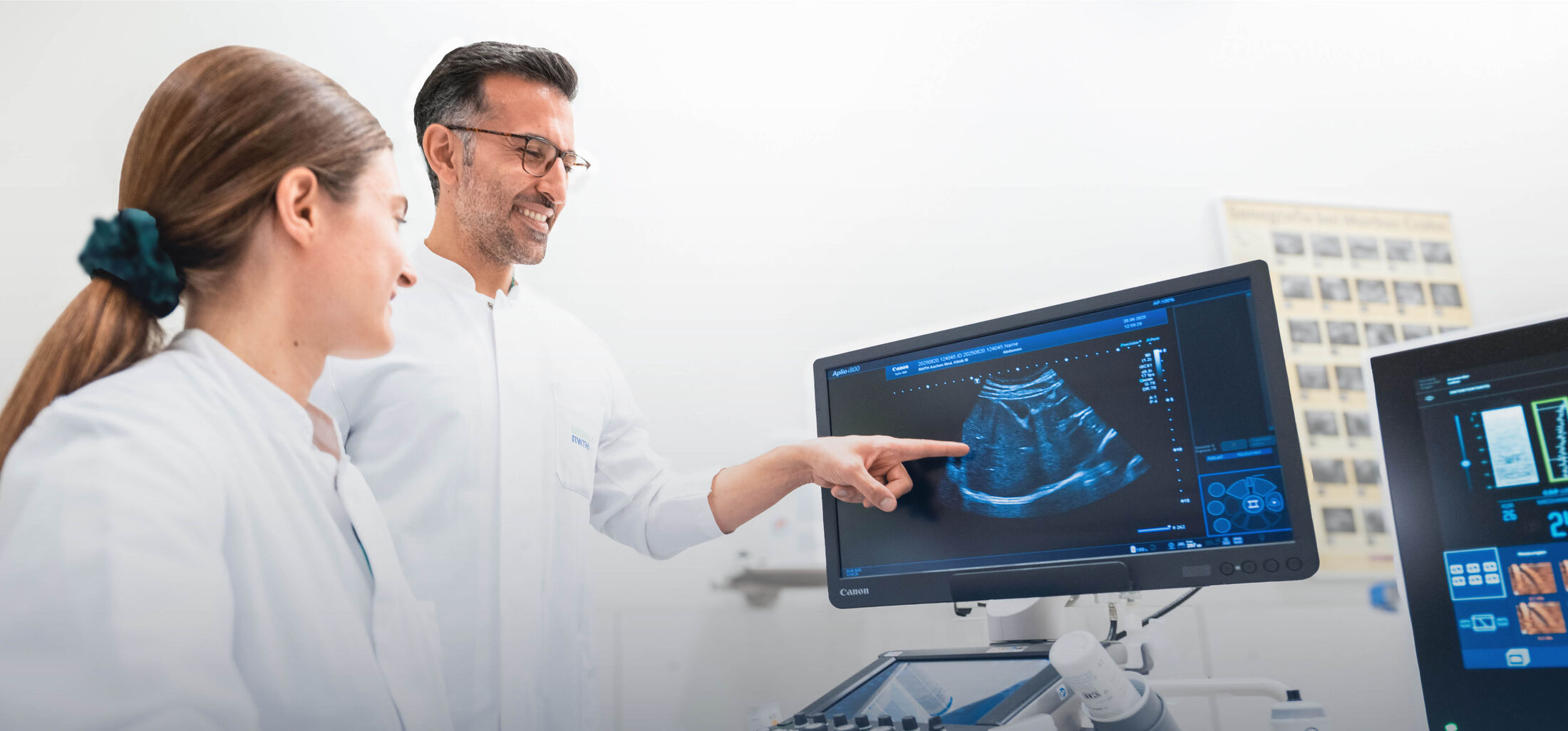

In addition to first-class equipment, our interdisciplinary ultrasound unit has several experienced and certified physicians. We can therefore offer you all standard ultrasound procedures at the highest quality standard.
We would like to briefly introduce, in plain language, the main procedures we routinely offer. During an outpatient or inpatient stay, we will gladly explain the background of the procedures relevant to you and their advantages and disadvantages for your individual situation.
Sonography (commonly called ultrasound) uses ultrasound waves and their reflection to generate moving images. This allows internal organs to be visualized in detail and without radiation (and thus without side effects). Because the examination happens "live," findings can be communicated immediately and discussed. These advantages also make it ideal for side-effect-free follow-up monitoring.
Sonography has undergone an impressive development over the past decades. Please feel free to take a look at the further explanatory texts on this topic.
Examination of the dynamic image of internal organs is the cornerstone of every ultrasound examination. The examination is performed lying down on a padded table; positioning maneuvers are sometimes helpful for further clarification. To achieve the best possible image, we occasionally need your assistance, e.g. through breathing maneuvers. The examination takes only a few minutes, is painless, and causes no radiation exposure whatsoever (unlike, for example, X-ray examinations).
In our interdisciplinary ultrasound center, we offer sonographic diagnostics of all internal abdominal organs as well as the thyroid, using the special/innovative techniques described below.
In some cases, you must be fasting in order to allow for better diagnostics. Please take a look at the further explanatory texts on this topic, or contact us if you have any questions.
For specific questions, the dynamic ultrasound image is supplemented by velocity measurements, so-called color Doppler sonography (also called "duplex"). This allows precise examination of blood flow in vessels. We also have additional contrast-free techniques available (e.g. advanced dynamic flow (ADF) or superb micro-vascular imaging (SMI)). This examination is also painless and risk-free.
By adding contrast agents into the vessels, ultrasound can also show the blood supply of certain organ abnormalities comparably well to computed tomography (CT) or magnetic resonance imaging (MRI). This procedure is called contrast-enhanced ultrasound, or, from the English term, "CEUS" (contrast-enhanced ultrasound). The advantage here is that the ultrasound contrast agent is virtually risk-free (e.g. no kidney damage). This procedure is particularly valuable in liver tumor diagnostics. Prior to the procedure and the placement of an intravenous indwelling cannula, a consent discussion takes place. The contrast agent is administered intravenously and is painless.
With the help of measurements of tissue stiffness (elastography in the narrower sense) and, for the liver, also the attenuation of the ultrasound signal (the so-called "controlled attenuation parameter" (CAP)), important information about organ changes can be determined. This allows for a reliable assessment of whether an organ such as the liver shows scarring (via the measurement of liver stiffness) or fatty liverdisease (via the measurement of CAP). In addition to so-called transient elastography (FibroScan® by Echosens), 2D shear-wave elastography by Canon is also available to us. The examination is painless and, just like ultrasound, carries no risks. It is performed lying down, following the standard sonography examination. Thanks to elastography, many patients can now be spared a tissue sample, although this remains essential in selected cases.
Important: For a reliable result, you should appear fasting (no fluids and no food for 4 hours).
Taking a tissue sample (biopsy) for microscopic histological analysis is often crucial for a definitive diagnosis. Such an organ puncture is usually quick and low-risk under ultrasound guidance and local anesthesia. During the preliminary consultation, a physician experienced in ultrasound will examine you and inform you of your individual benefit and risk. Usually a brief period of monitoring on our "same-day intervention" unit follows, so you can go home the same day; in some cases an overnight stay is necessary.
So-called minimally invasive therapies offer, through a small or minor procedure, a comparable benefit with lower risk for selected conditions. Such therapies can be performed under local anesthesia or general anesthesia. During the preliminary consultation an ultrasound-experienced physician will examine you and inform you of your individual benefit and risk. If several procedures are possible, this will also be discussed with you. Usually a period of inpatient monitoring follows before you are discharged home.
Before the Examination
We are happy to offer you our entire ultrasound repertoire, which meets the highest quality standards. If your treating physician requests further diagnostics, or if you would like a second opinion yourself, we will be glad to examine you and share our assessment with both you and your treating physician.
Depending on the question or need, we also apply special and innovative techniques, at no additional cost to you. In addition to a "normal ultrasound" (native sonography), we therefore also perform a special imaging of the vessels (color Doppler sonography). Should a more precise understanding of structures be required, we perform a contrast-enhanced sonography following a corresponding consent discussion with you. In the case of acute or chronic liver disease, we can, in addition to native and color Doppler sonography, also measure tissue stiffness or fatty infiltration using elastography (FibroScan®).
During the examination, we explain the findings to you and let you know whether any recommendations for further action arise from them. The findings, along with the images, are handed to you directly after the examination. This gives you the opportunity to discuss them with your family doctor or your referring physician.
Should you or your referring physician have a specific question that needs to be answered, it would be helpful if you brought prior reports and current lab values with you. It is also important that you appear fasting (no eating or drinking for four hours).
We are happy to remain available afterward for any questions or changes in findings. Please understand that, in the case of a complex issue and/or insufficient prior findings, an additional presentation at one of our specialist consultations may become necessary so that we can give you a reliable assessment.
Please understand that our capacity is limited and that waiting times for appointments may occur. A prompt examination is only possible at the request of a physician with a corresponding referral.
What should you note before an ultrasound examination?
To optimize image quality, it would be advisable to remain fasting for four hours before your appointment, meaning you should not eat or drink anything for the four hours prior to the examination. In particular, carbonated beverages should be avoided during this period. If this is possible for you, an examination can also be performed without prior fasting (however, this is mandatory for the reliable performance of an elastography (FibroScan)!). It would at least be advisable to avoid gas-inducing foods on the day before the examination, as the quality of the examination can be impaired by air in the abdomen. A visit to the toilet is not necessary before the examination.
Please also remember to bring a referral form. Should you or your referring physician have a specific question that needs to be answered, it would be helpful if you brought prior findings and current lab values with you.
What should you note before an ultrasound-guided procedure?
Before performing an ultrasound-guided procedure, informed consent about potential risks and complications is essential. A consent discussion must take place at least the day before the procedure. Your treating physician will therefore hold a consent discussion with you at an agreed appointment. Please inform us if you have an allergy or hypersensitivity reaction to medications, adhesive plasters, local anesthetics, or similar, or if you suffer from a serious cardiovascular or pulmonary disease or an infectious disease. During the consent discussion, you will be informed not only about the general course of the examination but also about the precise reason for it in your individual case, as well as the risks and side effects. Please understand that such an examination cannot be performed without adequate consent having been given.
Please bring a referral form issued by your family doctor on the day of your examination, as well as the completed and signed consent forms. Furthermore, we require lab values (complete blood count, coagulation) that are no older than 4 weeks. For your protection and for better visualization, the procedure is performed while you are fasting. Your last food intake should therefore have been at least 6 hours prior. Please also refrain from consuming fluids and nicotine on the day of the examination. You should clarify with your physician to what extent your regularly taken medications should also be omitted on the morning of the examination day (blood thinners in particular). It may be necessary to discontinue one or all medications that affect blood clotting 5-7 days beforehand (e.g. ASA, Marcumar, Iscover, Plavix, Xarelto, Eliquis, etc.). Please discuss this with your treating physician.
If you are traveling by your own car, you should bear in mind that you may be given pain medication that impairs your fitness to drive. Following the administration of such medication, you may not actively participate in road traffic for 12-24 hours. You should therefore consider arranging for an accompanying person for your return journey, if applicable!

